42 heart structure with labels
› heart-healthHeart Health | Heart Attack Prevention | Bayer® Aspirin TO HELP PREVENT ANOTHER HEART ATTACK. A doctor-directed aspirin regimen helps keep your blood flowing. Along with other heart-healthy choices, it can reduce your risk of having another heart attack. Learn About Aspirin's Benefits. Aspirin is not appropriate for everyone, so be sure to talk to your doctor before you begin an aspirin regimen. Understanding an ECG | ECG Interpretation | Geeky Medics An ECG lead is a graphical representation of the heart's electrical activity which is calculated by analysing data from several ECG electrodes. A 12-lead ECG records 12 leads, producing 12 separate graphs on a piece of ECG paper. Only 10 physical electrodes are attached to the patient, to generate the 12 leads. Electrodes
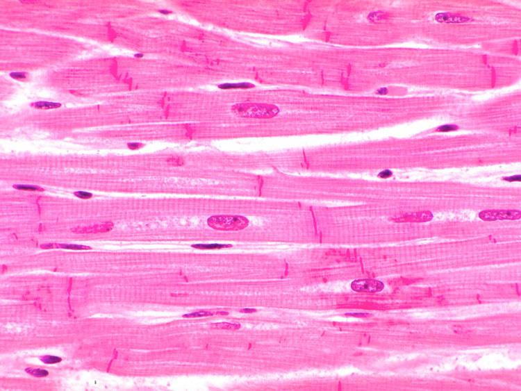
Heart (right and left atrium): Anatomy and function | Kenhub The heart contains three main layers of tissue. The innermost layer is known as the endocardium and the outermost layer is the epicardium. Between those two layers is a thick layer of specialized (i.e. cardiomyocytes) known as the myocardium. The thickness of the myocardium varies between regions of the heart.

Heart structure with labels
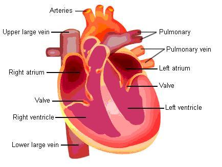
Heart & Circulatory System Diagram, Parts & Function, For Kids The heart comprises four chambers. Right and left atria are the two upper chambers. These chambers are separated by a wall called the interatrial septum. Right and left ventricles are the two lower chambers. The interventricular septum separates these chambers. Atrioventricular valves separate the atria and ventricles on each side of the heart. Diagram of Human Heart and Blood Circulation in It | New ... A heart diagram labeled will provide plenty of information about the structure of your heart, including the wall of your heart. The wall of the heart has three different layers, such as the Myocardium, the Epicardium, and the Endocardium. Here's more about these three layers. Epicardium Identify Various Parts Of A Human Heart: Trivia Quiz ... The heart is the most important organ in the body. It is in charge of keeping the processes within the body moving by facilitating the transfer of blood throughout the body. The quiz below is to test out interesting facts you may know about the heart. Give it a try and good luck. Questions and Answers 1. What is F? 2. What is K? 3. What is C? 4.
Heart structure with labels. Heart Labeling Quiz: How Much You Know About Heart ... Here is a Heart labeling quiz for you. The human heart is a vital organ for every human. The more healthy your heart is, the longer the chances you have of surviving, so you better take care of it. Take the following quiz to know how much you know about your heart. Questions and Answers 1. What is #1? 2. What is #2? 3. What is #3? 4. What is #4? Heart - Wikipedia In humans, other mammals, and birds, the heart is divided into four chambers: upper left and right atria and lower left and right ventricles. Commonly the right atrium and ventricle are referred together as the right heart and their left counterparts as the left heart. byjus.com › biology › human-heartHuman Heart - Anatomy, Functions and Facts about Heart The external structure of the heart has many blood vessels that form a network, with other major vessels emerging from within the structure. The blood vessels typically comprise the following: Veins supply deoxygenated blood to the heart via inferior and superior vena cava, and it eventually drains into the right atrium. Circulatory System Diagram - New Health Advisor Circulatory System Diagram. The circulatory system is the most vital systems of your body that is required for the optimal distribution of oxygenated blood to all the body organs and tissues. A fully functional circulatory system aims to maintain adequate concentration of oxygen in the biological tissues to ensure longevity and health.
Anatomy of the heart and coronary arteries (coronary CT ... 1. Basal anterior 10. Mid inferior 11. Mid inferolateral 12. Mid anterolateral 13. Apical anterior 14. Apical septal 15. Apical inferior 16. Apical lateral 17. Apex 2. Basal anteroseptal 3. Basal inferoseptal 4. Basal inferior 5. Basal inferolateral 6. Basal anterolateral 7. Mid anterior 8. Mid anteroseptal 9. Mid inferoseptal Acute marginal (AM) › en › healthy-livingCarbohydrates | American Heart Association Carbohydrates are either called simple or complex, depending on the food’s chemical structure and how quickly the sugar is digested and absorbed. The type of carbohydrates that you eat makes a difference – Foods that contain high amounts of simple sugars, especially fructose raise triglyceride levels. Layers of the heart: Epicardium, myocardium, endocardium ... The endocardium is the innermost layer of the heart. It lines the inner surfaces of the heart chambers, including the heart valves. The endocardium has two layers. The inner layer lines the heart chambers and is made of endothelial cells. Body Cavities Labeled: Organs, Membranes, Definitions ... Body cavities along with their organs and membranes simplified! Labeled diagrams, definitions, and lateral views included! High-yield flow chart and table of the dorsal, ventral, cranial, spinal, thoracic, pleural, pericardial, abdominal, and pelvic cavities!
How the Heart Works: Diagram, Anatomy, Blood Flow The heart is located under the rib cage -- 2/3 of it is to the left of your breastbone (sternum) -- and between your lungs and above the diaphragm. The heart is about the size of a closed fist, weighs about 10.5 ounces, and is somewhat cone-shaped. It is covered by a sack termed the pericardium or pericardial sack. Heart: illustrated anatomy - e-Anatomy 1. Basal anterior 10 - Second diagonal 10. Mid inferior 10a - Second diagonal a 11 - Proximal circumflex 11. Mid inferolateral 12 - Intermediate/anterolateral 12. Mid anterolateral 12a - Obtuse marginal a 12b - Obtuse marginal b 13 - Distal circumflex 13. Apical anterior 14 - Left posterolateral 14. Apical septal 14a - Left posterolateral a 20 Heart and Circulatory System Activities For Kids ... A healthy heart needs exercise too. Create a tic-tac-toe board with the free printables at the link, then toss a bean bag (bonus points for making heart-shaped ones!) to see which exercise you'll do next. Learn more: Make and Takes. 20. Host a Jump Rope for Heart event Know the Structures and Functions about Your Heart | New ... Heart structure and function are closely related, as described below: 1. The Pericardium This is a fibrous covering that wraps around the heart and holds it in place. This special membrane also contains a fluid which lubricates the heart in the pericardial space or cavity to prevent friction.
Know the Structures and Functions about Your Heart | New Health Advisor
› indexCells and cell structure quiz questions - Footprints-Science ... Cell structure Quiz Cell division Quiz Transport in cells Quiz Digestive system Quiz Heart and blood Quiz Health issues Quiz Plant tissues, organs and systems Quiz Communicable diseases Quiz Drugs Quiz Plant disease Quiz Photosynthesis Quiz Respiration Quiz Homeostasis Quiz Nervous system Quiz Hormones Quiz Reproduction Quiz Variation and ...

Pin on microscope slides
Cardiovascular system: Diagrams, quizzes, free ... - Kenhub Comprised of the heart, blood vessels and the blood itself, it is divided into two loops which both begin in the heart. The pulmonary circuit is responsible for exchanging blood between the heart and lungs for oxygenation, while the systemic circuit directs blood to the other tissues of the body.
Tissues Flashcards | Easy Notecards
Anatomy, Thorax, Heart Coronary Arteries - NCBI Bookshelf The coronary arteries run along the coronary sulcus of the myocardium of the heart. Their main function is to supply blood to the heart. This is a crucial function for myocardial function and subsequently homeostasis of the body. The arrangement of coronary arteries varies among people significantly.

The human egg cell explained for egg donors | Altrui
› design-templates › printHeart Diagram – 15+ Free Printable Word, Excel, EPS, PSD ... Teachers and students use the heart diagram, in biological science, to study the structure and functions of a human being’s heart. Friends and colleagues on the other hand may find this diagram template useful when it comes to sending special, personalized gifts to their family members and significant others. Download the template today, and ...
CDC - Congenital Heart Defects, Atrial Septal Defect, Graphic- NCBDDD
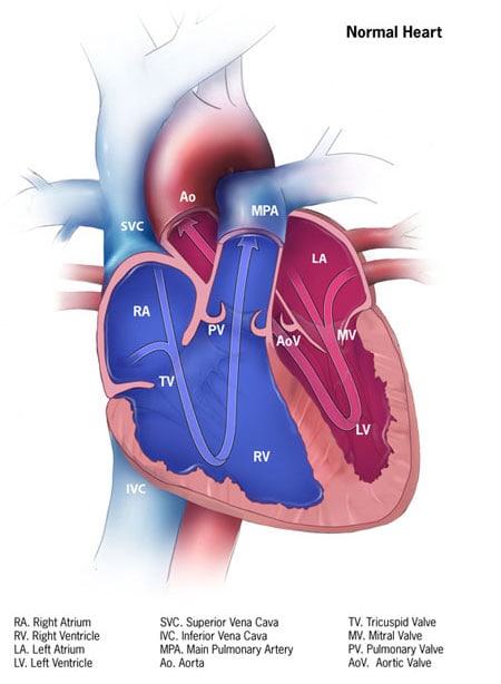
Human heart: Anatomy, function & facts | Live Science The physiology of the heart basically comes down to "structure, electricity and plumbing," Phillips told Live Science. The human heart has four chambers: two upper chambers (the atria) and two ...

how to draw label diagram of heart - Science - Life Processes - 12575 | Meritnation.com
Heart Drawing With Label : Human Heart Diagram Images ... Heart Drawing With Label : Human Heart Diagram Images Stock Photos Vectors Shutterstock. Internal structure of human heart shows four chambers viz. Download this sketch of human heart anatomy with hand written labels vector illustration now. Two atria and two ventricles and couple of blood vessels opening into them.
Heart Labeling (Internal)
Heart Simple Drawing With Labels - How To Draw Human Heart ... External structure of human heart shows its conical shape with apex facing downwards. And search more of istock's library of . You can learn diagram of heart with labels and easy simple heart anatomy with heart . Internal structure of human heart shows four chambers viz. Label the parts neatly as shown.

Heart Diagrams for Labeling and Coloring, With Reference Chart and Summary | Heart diagram ...
Mnemonics for Heart Anatomy and Physiology (Video) A heart block is an abnormal heart rhythm known as an arrhythmia and can occur anywhere in the specialized conduction system of the heart. The electrical signals telling the heart to contract are partially or totally blocked between the atria and ventricles. Therefore, it is called an atrioventricular, or AV block.

Labelled Heart by abpischools - Teaching Resources - Tes
The Three Layers of the Heart Wall - Video & Lesson ... The epicardium, also known as the visceral pericardium, is a layer of the parietal pericardium that reflects down and directly adheres to the heart tissue. This means that both the visceral...
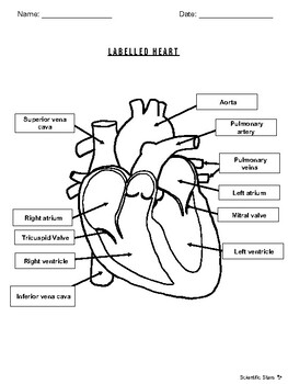
Labelled Heart by Scientific Stars | Teachers Pay Teachers
Circulatory System Anatomy, Diagram, & Function There are three layers of the heart wall. The epicardium is the heart wall's outer layer, the myocardium is the middle — and muscular — layer, and the endocardium is the heart's innermost layer....
Cardiac Physiology
How the Heart Works - The Heart | NHLBI, NIH The heart is an organ about the size of your fist that pumps blood through your body. It is made up of multiple layers of tissue. Your heart is at the center of your circulatory system. This system is a network of blood vessels, such as arteries, veins, and capillaries, that carries blood to and from all areas of your body.
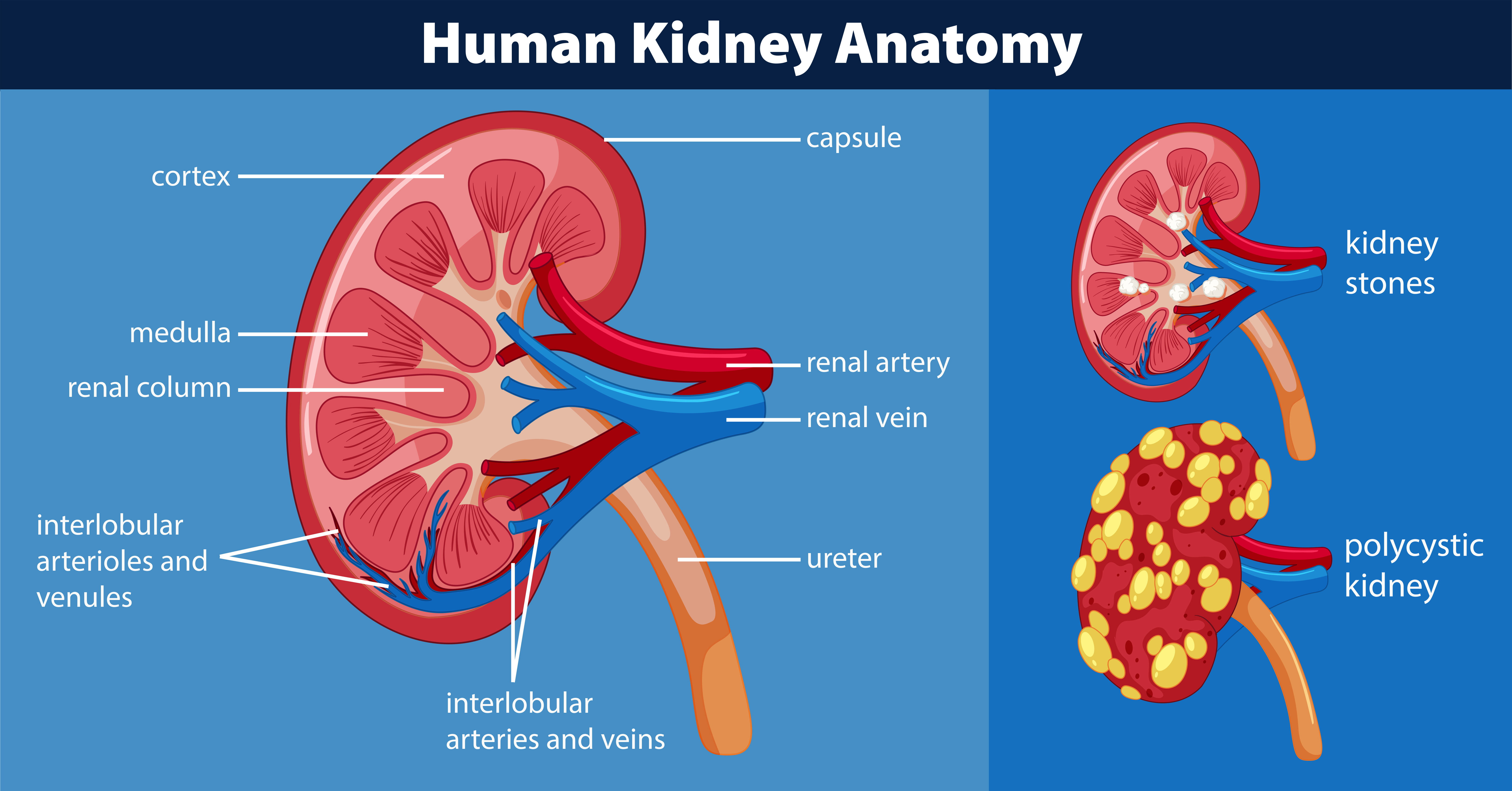
Human kidney anatomy diagram 446409 Vector Art at Vecteezy
Diagrams, quizzes and worksheets of the heart | Kenhub Labeled heart diagrams Take a look at our labeled heart diagrams (see below) to get an overview of all of the parts of the heart. Once you're feeling confident, you can test yourself using the unlabeled diagrams of the parts of the heart below. Labeled heart diagram showing the heart from anterior Unlabeled heart diagrams (free download!)

Simplified Heart Labeled Decal | Shop Fathead Anatomical Images Graphics
Heart histology: Cells and layers - Kenhub The heart contains three basic layers similar to those seen in arteries and veins. The outermost layer is the epicardium, which is derived from the proepicardium (from the septum transversum). The middle layer is the myocardium, and the innermost layer is the endocardium, which originated from mesothelial cells of the outflow tract.

Parts Of Heart Diagram Stock Illustration - Download Image Now - iStock
Lab 2: Anatomy of the Heart - Anatomy & Physiology: BIO ... It looks like you're using Internet Explorer 11 or older. This website works best with modern browsers such as the latest versions of Chrome, Firefox, Safari, and Edge.
![Untitled Document [www.bio.sunyorange.edu]](http://www.bio.sunyorange.edu/updated2/THINKING_EVOLUTION/physiology1/heart/c_chambers.jpg)
Untitled Document [www.bio.sunyorange.edu]
Anatomy of the Heart: Blood Flow and Parts - Video ... Four Chambers The ventricles pump blood out of the heart. Your heart has four hollow chambers. The two chambers on top are called the atria. These are the heart chambers that receive blood...
Post a Comment for "42 heart structure with labels"