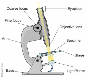
45 microscope diagram without labels
› products › microscopeMicroscope Objective Lens | Products | Leica Microsystems The objective lens is a critical part of the microscope optics. The microscope objective is positioned near the sample, specimen, or object being observed. It has a very important role in imaging, as it forms the first magnified image of the sample. The numerical aperture (NA) of the objective indicates its ability to gather light and largely determines the microscope’s resolution, the ... › category › newsNews Archives | Hollywood.com Travel through time by exploring Hollywood.com's entertainment news archives, with 30+ years of entertainment news content.
Compound Microscope Parts, Diagram Definition, Application, Working ... Definition of a Compound Microscope and uses. parts of a compound microscope and application. compound microscope labeled diagram. MN . Generic selectors. ... adhesive labels are stuck to the base and sides of the microscope. ... It is not recommended to use oil immersion lenses without oil. Use of Compound microscope. Compound Microscopes used ...

Microscope diagram without labels
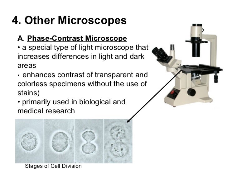
Light Microscope (Theory) - Amrita Vishwa Vidyapeetham Microscope can be separated into optical theory microscopes (Light microscope), electron microscopes (eg.TEM, SEM) and scanning probe microscopes. (eg.AFM, PSTM). Optical microscopes function on the basis of optical theory of lenses by which it can magnifies the image obtained by the movement of a wave through the sample. Gram Stain Technique - Amrita Vishwa Vidyapeetham You may also label the slide with the initials of the name of the organism on the edge of the slide. Care should be taken that the label should not be in contact with the staining reagents. ... Do not ever observe at a specimen at 100X without oil. While focusing the microscope, glass slides should be handled carefully to avoid the chance of ... Plant Cell Parts And Functions With Picture Plant Cell Diagram 1 Cell Wall It is the outermost protective layer of a plant cell having a thickness of 20-80 nm. Controls passage of materials in and out of the cell cytoplasm everything inside of the cell membrane except for the nucleus light green nucleus control center of the cell. Parts of a plant cell.
Microscope diagram without labels. EOF › questions-and-answers › learningAnswered: Learning through Art: Water Molecules… | bartleby Transcribed Image Text: › en › microscopeFluorescence Resonance Energy Transfer (FRET) Microscopy Presented in Figure 3 is a Jablonski diagram illustrating the coupled transitions involved between the donor emission and acceptor absorbance in fluorescence resonance energy transfer. Absorption and emission transitions are represented by straight vertical arrows (green and red, respectively), while vibrational relaxation is indicated by wavy ... Terahertz thermal curve analysis for label-free identification of ... We developed a label-free identification tool for individual pathogens, such as bacteria, by performing THz thermal curve analysis, as schematically illustrated in Fig. 1a. We monitored the...
Plant Cell: Diagram, Types and Functions - Embibe Exams Despite the fact that plant and animal cells are both eukaryotic and share a few cell organelles, plant cells perform different roles than animal cells. When the cells are inspected under an electron microscope, some of these changes become obvious. In this article read more about Plant Cell, Diagram, Functions, and Types. › topics › medicine-andConfocal Microscopy - an overview | ScienceDirect Topics A standards document, which describes confocal microscopy and its influence quantities, has recently completed an ISO ballot as a final draft international standard (ISO FDIS 25178-607, 2018). A schematic diagram of a typical confocal microscope is shown in Fig. 15.1 (ASME B46-2009, 2010; Weller et al., 2012). Most examples of this method rely ... en.wikipedia.org › wiki › WavelengthWavelength - Wikipedia In physics, the wavelength is the spatial period of a periodic wave—the distance over which the wave's shape repeats. It is the distance between consecutive corresponding points of the same phase on the wave, such as two adjacent crests, troughs, or zero crossings, and is a characteristic of both traveling waves and standing waves, as well as other spatial wave patterns. pages.zeiss.com › rs › 896-XMS-794Principles of Fluorescence and Fluorescence Microscopy fies the principle of the fluorescence microscope — without the light-filtering abilities of the purple glass window and the glass of white wine, Stokes would not have been able to observe any fluorescence at all. Using Stokes’ observation and the green fluorescent protein (GFP) as examples, this article will explain
Plant Cell Parts And Functions With Picture Plant Cell Diagram 1 Cell Wall It is the outermost protective layer of a plant cell having a thickness of 20-80 nm. Controls passage of materials in and out of the cell cytoplasm everything inside of the cell membrane except for the nucleus light green nucleus control center of the cell. Parts of a plant cell. Gram Stain Technique - Amrita Vishwa Vidyapeetham You may also label the slide with the initials of the name of the organism on the edge of the slide. Care should be taken that the label should not be in contact with the staining reagents. ... Do not ever observe at a specimen at 100X without oil. While focusing the microscope, glass slides should be handled carefully to avoid the chance of ... Light Microscope (Theory) - Amrita Vishwa Vidyapeetham Microscope can be separated into optical theory microscopes (Light microscope), electron microscopes (eg.TEM, SEM) and scanning probe microscopes. (eg.AFM, PSTM). Optical microscopes function on the basis of optical theory of lenses by which it can magnifies the image obtained by the movement of a wave through the sample.
Biology: January 2011

31 Label The Indicated Parts Of The Microscope - Labels For Your Ideas
Search in gallery
![Cikgu Naza: [Biology Form 4] Animal Cell & Plant Cell](https://blogger.googleusercontent.com/img/b/R29vZ2xl/AVvXsEhgZk71NMh_vLioF6_4OGNAXrAtBvN-Cc4pgR4ctFfp8rqREiuSK5iHyWOLOEU-qZ6x5KXM46f5jy5IEoJYGTp3PuYr3RsgJxnGO1rOrmnxZhmoc_VOFZqp5ZmFlLZpXjn4I4sddI-OgK-H/s400/plant+cell.JPG)
Cikgu Naza: [Biology Form 4] Animal Cell & Plant Cell

BIOLOGY ORDINARY LEVEL NOTES: CELL STRUCTURES

A schematic diagram of the light path in the ptychographic microscope... | Download Scientific ...

Prokaryotic cells (Prokaryotes): Definition, Structure, Parts, Examples and Diagram

Mr. Mak's Grade 8 Science Website
Topic 3 - Microscopes

Simple Microscope Labelled Diagram - Micropedia
Labeled-microscope-diagram

30 Label Diagram Of Microscope

A Study of the Microscope and its Functions With a Labeled Diagram | Teaching ideas
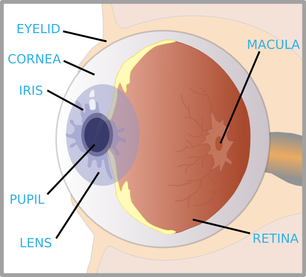
Eye With Labels Clip Art at Clker.com - vector clip art online, royalty free & public domain
Guide to Using a Microscope - Year 8 Portfolio
Post a Comment for "45 microscope diagram without labels"